Dávalos Lab
Dávalos Lab
News
People
Projects
Publications
Courses
Join
Contact
Photos
Archive
neocortex
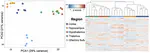
Histological and MRI brain atlas of the common shrew, Sorex araneus, with brain region-specific gene expression profiles
The common shrew,
Sorex araneus
, is a small mammal of growing interest in neuroscience research, as it exhibits dramatic and reversible …
Cecilia Baldoni
,
William R. Thomas
,
Dominik von Elverfeldt
,
Marco Reisert
,
Javier Lázaro
,
Marion Muturi
,
Liliana M. Dávalos
,
John D. Nieland
,
Dina K.N. Dechmann
PDF
Cite
Project
Source Document
DOI
Cite
×